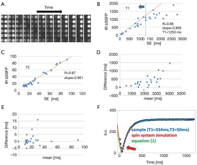
Figure 1.
(A) In vitro cross-sectional images from an IR-bSSFP acquisition. Images are shown every 56 msec. Varying signal intensities due to different T1 and T2 values can be appreciated; (B) Comparison between T1 values obtained with TSE and IR-bSSFP. The straight line corresponds to the function f(x)=x. The R value was calculated with T1 values lower than 1,250 ms (determined with TSE, in graph corresponding to data points left of blue line as indicated by blue arrow); (C) Comparison between T2 values obtained with TSE and IR-bSSFP. The straight line corresponds to the function f(x)=x. R value was calculated with all T2 values shown; (D) Bland-Altman plot for values in C (T1) illustrating the systematic underestimation of the T1 determined with IR-bSSFP for larger values; (E) Bland-Altman plot for values in D (T2). Good agreement can be observed without systematic deviations; (F) Results of spin system magnetization (red line), equation (1, green line) and in vitro measurement (blue dots) for T1=344 ms and T2=50 ms (from TSE). Good overall agreement can be appreciated with largest deviations occurring where the longitudinal magnetization changes sign (arrow). Abbreviation: IR-bSSFP, inversion recovery balanced steady state free precession; TSE, turbo spin-echo.

