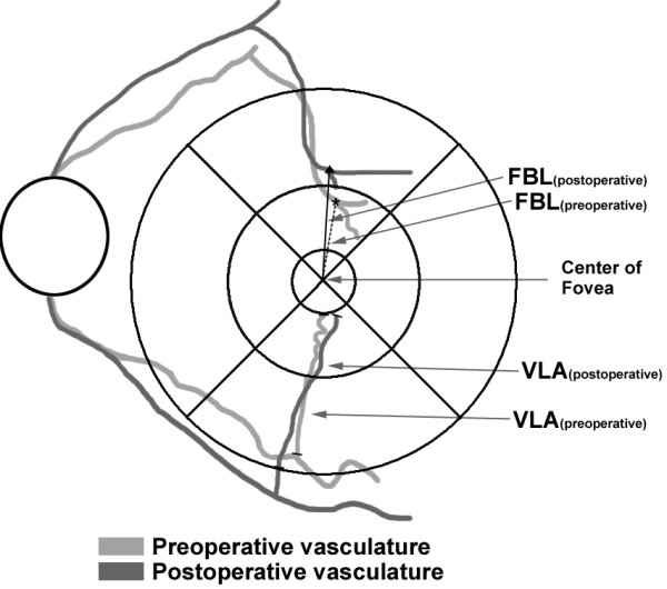
Figure 1.

A schematic showing the two features of change in the retinal surface. (Upper quadrant) Length from the foveola to the vessel branching point (FBL) was calculated after selection of five points of the vessel branching points located at the superior and the inferior quadrant, in order of proximity to foveola. (Lower quadrant) Vessel segment length in area (VLA) was defined as the vessel segment length for radial vessels included in each quadrant consisting of a 1 mm and 6 mm diameter ring. Only vessel segments included between the 1 mm ring and the 6 mm ring was measured. The VLA was measured by selecting 3–5 radial arterioles or venules in each quadrant. FBL and VLA were calculated using infrared fundus photographs and image processing software (ImageJ).
