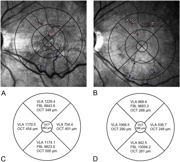
Figure 2.
Comparison of pre- and postoperative topographic parameters in the same patient with ERM. (A) Infrared fundus photograph (overlaid with Early Treatment Diabetic Retinopathy Study [ETDRS] subfield) shows tortuous macular vessels preoperatively. Points a to j indicate the vessel branching points located at the superior and inferior areas in order of proximity to fovea (B) Postoperative infrared fundus photograph shows release of macular contraction. Points a* to j* indicate displaced branching points after surgery. (C) Actual preoperative measurement values of the vessel segment length in area (VLA), the length from the foveal center to the vessel branching point (FBL), and optical coherence tomography (OCT) are shown in each area. (D) Postoperative VLA, FBL, and OCT values are shown.

