
Figure 1.
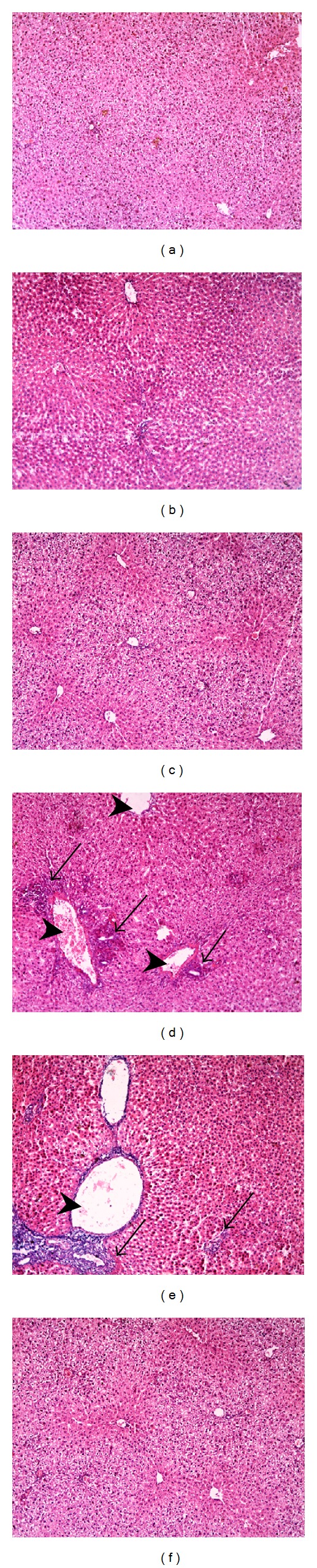
Effect of fenofibrate (FEN) and pioglitazone (PIO) on liver histopathological profile in cyclophosphamide (CP)-treated rats. Representative photomicrographs of liver from: ((a), (b), and (c)) control and FEN and PIO groups, respectively, showing no pathological changes in hepatocytes, (d) CP-treated group presenting with loss of normal hepatic architecture, congested dilated central vein, inflammatory cellular infiltration, and perivenular hepatocytic necrosis, (e) CP/FEN group showing congested dilated central veins with focal inflammatory cellular infiltration and degenerative necrotic cells, and (f) CP/PIO group demonstrating normal liver histology. Arrowhead: dilated central vein, black arrow: inflammatory cellular infiltration (×100). The histological changes were scored, and results are expressed in Table 1.