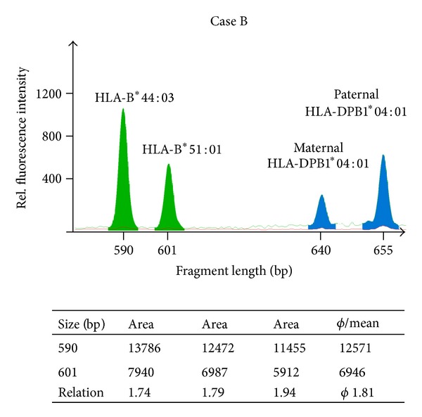
Figure 4.

RFQ of HLA-B. Complement-dependent cytotoxicity donor-depleted cells of the second patient (Case B) were analyzed by RFQ which has taken advantage of an HLA-B length polymorphism in intron 4. The HLA-B fragments shown here (green filled peaks) have a size of 590 bp (B*44:03) and 601 bp (B*51:01). The fragments were amplified using the primers 5Bin3-389cons (TCC AGY ACT TCT GAG TCA CTT TAC) and 5′ Nulight547-labeled 3Bin4-56 (CCT GAC CCT GCT GAA GGG CTC C). The table below the figure shows the results of three independent experiments, which give an average allele measure of 1.82. These data are in good agreement with the HLA-DPB1 results. Nearly 50% of the leukemic cells also lacked the HLA-B region. Thus, both the HLA-DPB1 and the HLA-B locus revealed repression of the maternal-derived allele and HLA-B*51:01 was underrepresented. The HLA-DPB1 amplification products are also depicted (blue filled peaks).
