Fig. 6.

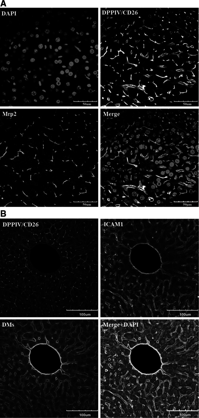
Validation of the DPPIV/CD26 and donkey anti-mouse IgG antibodies. a DPPIV/CD26 co-localizes with Mrp2. However, Mrp2 exclusively stains the bile canaliculi, whereas DPPIV/CD26 is also visible in the sinusoidal endothelial cells. The scale bars are 50 μm. b In architectural staining, donkey anti-mouse IgG (DMs) is used to visualize the sinusoidal endothelial cells. Co-staining with DMs (red) and the endothelial marker ICAM1 (white) shows good co-localization. The scale bars are 100 μm. DMs donkey anti-mouse IgG, DPPIV dipeptidyl peptidase IV, ICAM1 intercellular adhesion molecule-1, Mrp2 multi-drug resistance-associated protein 2 (colour figure online)
