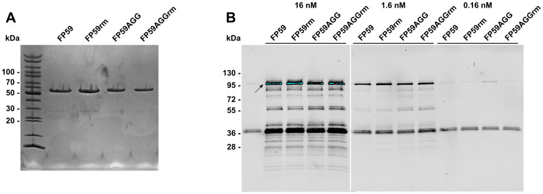
Figure 1. Analysis of FP59 fusion protein variants.
(A) Purified fusion proteins FP59 and FP59AGG were reductively methylated and analyzed by SDS-PAGE and Coomassie staining. This experiment was performed once to confirm the determined concentration of the proteins and to demonstrate purity of the samples. (B) All four FP59 variants were analyzed for the enzymatic activity of the PEIII domain by analysis of the ADP-ribosylation in vitro. Fusion proteins were incubated with partly purified eukaryotic elongation factor 2 from yeast in the presence of biotinylated NAD+ for 1 h at 37°C. Samples were analyzed by Western blotting and ADP-ribosylated eEF2 detected by streptavidin. This experiment was performed twice and one exemplary result is shown here.

