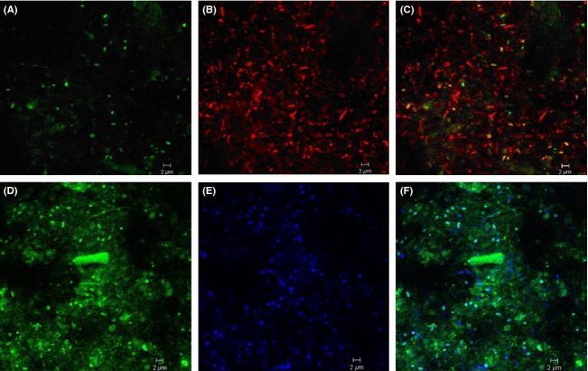
Figure 2.
In situ detection of the WWE1 group. Samples from mesophilic waste incubation hybridized with probe S-*-WWE1-408-a-A-19 in green (A) and Eub338mix in red (B), and superimposition of the two images (C). Samples from mesophilic cellulose incubation at day 14 hybridized with oligonucleotide probe S-*-WWE1-408-a-A-19 in green (D), and with probe Eub338mix in blue (E) and superimposition of the two images (F). The scale bar corresponds to 2 μm.

