
Figure 4.
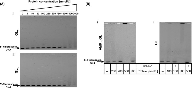
Electrophoretic mobility shift assays. (A) Performed with recombinant proteins corresponding to (i) the N-terminal – GLN and (ii) C-terminal – GLC regions of the catalytic domain of GL. Both proteins were able to shift 10 nmol/L of the 5′-fluorescein-labeled DNA fragment A, at high protein concentrations. (B) Competition electrophoretic mobility shift assays. All binding reactions were performed with 10 nmol L−1 of 5′-fluorescein-labeled fragment A, and ∼200-fold molar excess of unspecific DNA (low-molecular weight salmon sperm DNA, ssDNA) was added in the lanes specified. (i) AMR1–3GL protein, (ii) GL protein.