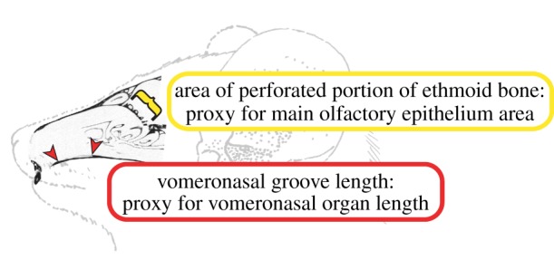
Figure 1.

Schematic diagram of the anatomical components of the mammalian olfactory chemosensory systems. The diagram shows a line drawing of Microcebus murinus with a CT scan (AMNH 174535) of the nasal fossa in the sagittal plane. The red arrows indicate the length of the VNG, which forms on the palatal portion of the maxilla from the articulation of the cartilaginous capsule surrounding the VNO. When data on VNO length were not available, we measured the VNG length as an osteological proxy. The yellow bracket indicates the perforated portion of the ethmoid bone, through which nerves projecting from the MOE pass and then connect to the MOB. We measured ethmoid area as a proxy for MOE area when data were not available in the literature. Line drawing adapted from Smith et al. [2]. (Online version in colour.)
