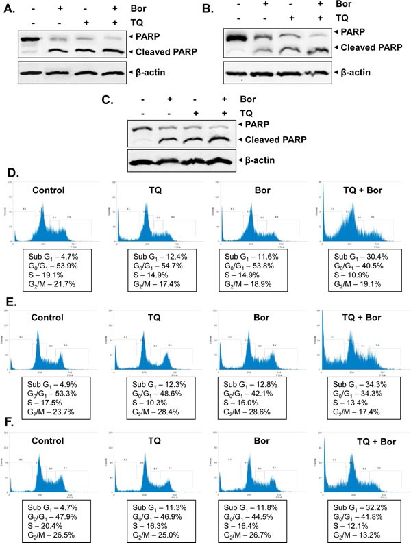
Fig 3. TQ potentiates the apoptotic effect of bortezomib in various MM cell lines.

A, H929 cells were treated with TQ (5 μM), bortezomib (20 nM) and a combination of both for 24 h at 37°C. Whole-cell extracts were prepared, separated on SDS-PAGE, and subjected to Western blot analysis using antibody against PARP. The same blots were stripped and reprobed with β-actin antibody to show equal protein loading. B, KMS-11 cells were treated with TQ (5 μM), bortezomib (20 nM) and a combination of both for 24 h at 37°C. Whole-cell extracts were prepared, separated on SDS-PAGE, and subjected to Western blot analysis using antibody against PARP. The same blots were stripped and reprobed with β-actin antibody to show equal protein loading. C, RPMI 8226 cells were treated with TQ (5 μM), bortezomib (20 nM) and a combination of both for 24 h at 37°C. Whole-cell extracts were prepared, separated on SDS-PAGE, and subjected to Western blot analysis using antibody against PARP. The same blots were stripped and reprobed with β-actin antibody to show equal protein loading. D, H929 cells were synchronized by serum starvation and then exposed to TQ (5 μM), bortezomib (20 nM) and a combination of both for 24 h at 37°C. The cells were then washed, fixed, stained with PI, and analyzed for DNA content by flow cytometry. E, KMS-11 cells were synchronized by serum starvation and then exposed to TQ (5 μM), bortezomib (20 nM) and a combination of both for 24 h at 37°C. The cells were then washed, fixed, stained with PI, and analyzed for DNA content by flow cytometry. F, RPMI 8226 cells were synchronized by serum starvation and then exposed to TQ (5 μM), bortezomib (20 nM) and a combination of both for 24 h at 37°C. The cells were then washed, fixed, stained with PI, and analyzed for DNA content by flow cytometry. Results typical of two independent experiments are shown.
