
Figure 7. Phosphorylation of AKT induced by actinomycin D (ActD).
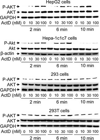
(A) 293, 293T, HepG2, and Hepa-1c1c7 cells were treated with ActD (0, 10, 30 and 100 nM) for 2, 6, and 10 minutes (min). The cells were then harvested, and cell lysates were analyzed by Western blotting using antibodies against Akt and anti-phospho-Akt (Ser473), GAPDH, and β-actin.