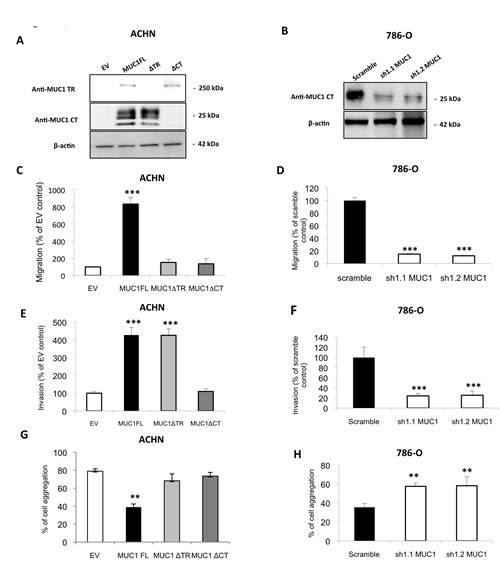
Figure 1. MUC1 increases migratory and invasive properties and decreases cell-cell interaction in ACHN and 786-O cells.

Western blotting were performed with anti–MUC1 targeting VNTR extracellular domain (M8) or cytoplasmic tail (Ab-5), and anti–β-actin antibodies on whole cell extracts obtained from (A) ACHN clones stably transfected with different expression vectors: MUC1-Full Length (MUC1FL), -deleted for its Tandem Repeat domain (MUC1ΔTR) or -deleted for its Cytoplasmic Tail (MUC1ΔCT) or an empty vector (EV) or (B) from 786-O clones stably transfected with a shRNA control (scramble) or with shRNA targeting MUC1 (sh1.1 and sh1.2). Cell migration of ACHN (C) and 786-O (D) clones was evaluated using 24-well migration chambers with 10% fetal calf serum as chemoattractant. The values obtained in EV-ACHN and scramble 786-O control cells were referred to as 100. The graphs show a percentage of control migration 24h after seeding. Values are means s.e.m (standard error mean) and represent five separate experiments (*** p<0.001). Cell invasion ((E) ACHN clones and (F) 786-O clones) was evaluated using 24-well Matrigel® invasion chambers with 10% fetal calf serum as chemoattractant. The values obtained in EV-ACHN and scramble 786-O control cells were referred to as 100. The graphs show a percentage of control invasion 24h after seeding. Values are means s.e.m and represent five separate experiments (*** p<0.001). ACHN (G) and 786-O (H) clones were seeded on agarose 0.8% under shaking. After 1h, aggregated and isolated cells were counted to determinate % of cell aggregation. Values are means s.e.m and represent at least three separate experiments (** p<0.01).
