Figure 1.
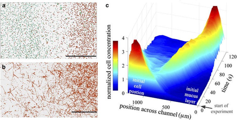
V. coralliilyticus is strongly attracted to coral mucus. (a) Positions and (b) trajectories of individual V. coralliilyticus cells exposed to a diffusing coral mucus gradient in a microfluidic channel (Supplementary Figure S1). A 400-μm thick layer of mucus, harvested from laboratory-cultured P. damicornis corals, was created in a microchannel (half of the layer is shown) and allowed to diffuse. The scale bars are 200 μm. In (a), cell positions at the start of the experiment and after two minutes are colored teal and red, respectively, and overlaid. In (b), trajectories acquired between 100 and 115 s after the start of the experiment are shown. The two panels show the strong shift in the cells' position and their intense accumulation into the mucus layer (the right side of the images). Also see Supplementary Movie S1. (c) The full time series of the spatial distribution of the pathogen population across the width of the microfluidic channel. Color and height both measure the local, instantaneous concentration of bacteria, normalized to a mean of one. Note the intense wave of bacteria actively migrating into the mucus layer.

