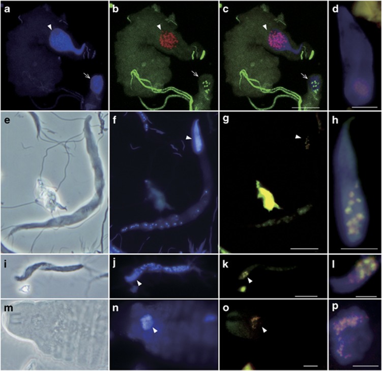
Figure 5.
Detection of verrucomicrobial symbionts in nuclei of (a–d) Pyrsonympha grandis, (e–h) Dinenympha exilis, (i–l) Dinenympha parva and (m–p) Teranympha mirabilis. (a, f, j, n) DAPI-stained images. (e, i, m) Phase contrast images. (b) Merged image of FISH analyses using the Texas red-labeled Verrucomicrobia-specific probe EUB338-III (red) and the 6FAM-labeled probe EUB338 specific to bacteria except Verrucomicrobia and several other taxa (green). (c) Merged image of a and b. (g, k, o) Merged images of FISH analyses using probe EUB338-III (red) and the 6FAM-labeled probe V-RsD37-130 specific to the majority of the termite-specific cluster in Figure 2 (green). (d, h, l, p) Magnified images of the DAPI-stained nuclei merged with FISH signals detected using probes EUB338-III (red) and V-RsD37-130 (green). Panel d was taken from a P. grandis cell that was different from the two cells in panels a–c. Arrowheads indicate the nuclei. Arrows in a–c indicate the Pyrsonympha nucleus that contained non-verrucomicrobial intranuclear symbionts. Panels a–c were observed under a laser scanning microscope. Bars represent 10 μm (c, g, k, o); 5 μm (d, h, l, p).

