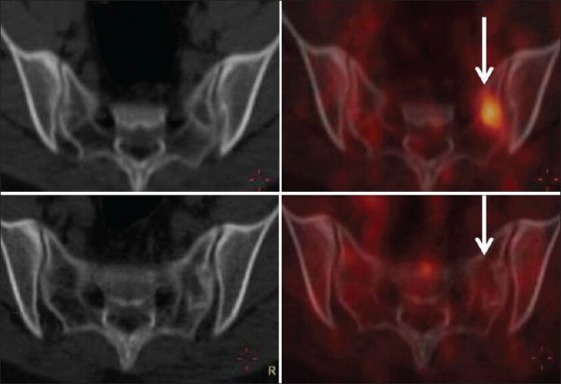
Figure 4.

Transaxial computed tomography (CT) and fused positron emission tomography (PET) CT images of a patient with primitive neuroectodermal tumor [patient no. 11, Table 3]. The upper panel shows no lesion in the sacrum on the CT image with a hypermetabolic marrow lesion in the left sacral ala in the fused PET CT image (arrow) (complete response and progressive metabolic disease-discordant result). The lower panel is follow up study showing minimal sclerosis in the left sacral ala in the CT image with no hypermetabolism in the fused PET CT image (arrow) suggesting response
