Figure 3.

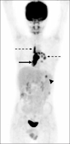
Maximum intensity projection image showing the presence of mediastinal and hilar lymphadenopathy (dotted arrows); splenic lesion (arrow head) and vertebral involvement (arrow) which could well have been mistaken for lymphoma recurrence

Maximum intensity projection image showing the presence of mediastinal and hilar lymphadenopathy (dotted arrows); splenic lesion (arrow head) and vertebral involvement (arrow) which could well have been mistaken for lymphoma recurrence