FIGURE 3.
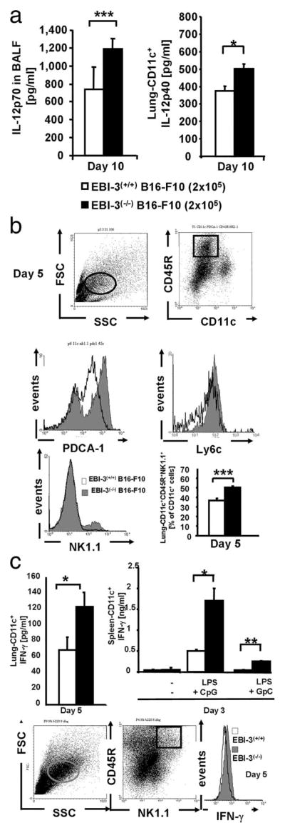
Analysis of immune cells and cytokines in the airways of EBI-3−/− mice developing lung melanoma. a, Left panel, Increased IL-12p70 levels were detected in the BALF from EBI-3−/− mice as compared with wild-type mice at day 10 after B16-F10 cell injection. Right panel, CD11c+ cells were isolated from the lungs of wild-type and EBI-3−/− mice 10 days after i.v. B16-F10 cell injection, and cultured overnight. The supernatant was then collected and analyzed by ELISA for IL-12p40 (n = 5). b, Increased number of CD11c+B220+ NK1.1+ cells in the lungs of EBI-3-deficient mice. Total cell suspensions from the lungs of EBI-3−/− and C57/BL6 wild-type mice were immunostained for the indicated markers and analyzed by FACS. The lung DCs were gated by following previously described methods (left upper panel). No significant difference in the total number of lung CD11c+cells was detected (1.860 ± 0.12 for EBI-3+/+ vs 2.58 ± 0.004 for EBI-3−/− CD11+ lung cells, respectively). The plasmacytoid DCs were identified as CD11c+CD45R (B220)+ PDCA-1+cells (26) (right upper panel and middle left panel, respectively) were found to be up-regulated in the lungs of EBI-3-deficient mice 5 days after tumor cell injection. Moreover, pDCs are Ly6c+. By gating on CD11c+B220+cells, we also found increased in CD11c+B220+Ly6c+ in the lung of EBI-3−/− mice. The IK-DC population was up-regulated in the lung of EBI-3−/− mice 5 days after tumor cell injection as compared with wild-type mice (right lower panel and middle right graph) n = 5. c, Left upper panel, Supernatants of lung CD11c+ cells from EBI-3−/− mice released increased amounts of IFN-γ 5 days after tumor cell injection as compared with those isolated from wild-type littermates. Lower panels, CD11c+ lung cells from EBI-3−/− and wild-type mice were immunostained for intracellular FACS analysis. On day 5 after tumor cell injection, EBI-3−/− lung cells expressing the markers for IK-DCs produced higher amounts of IFN-γ as compared with the wild-type littermates (right lower panel). Splenocytes derived from EBI-3−/− mice that were injected with B16-F10 cells 3 days before released increased amounts of IFN-γ as compared with those isolated from the wild-type littermates upon 24 h LPS (1 μg/ml) and CpG (10 μM) stimulation (upper right panel). All data represent mean values ± SEM of three independent experiments (n = 5). (*, p < 0.05; **, p < 0.01; ***, p < 0.001).

