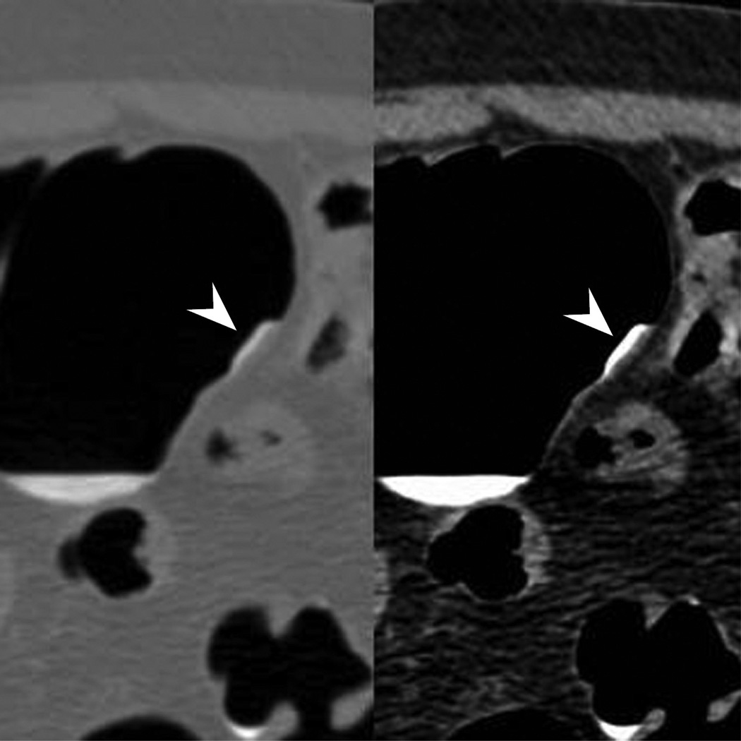
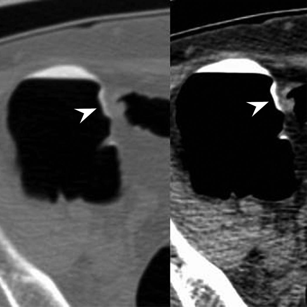
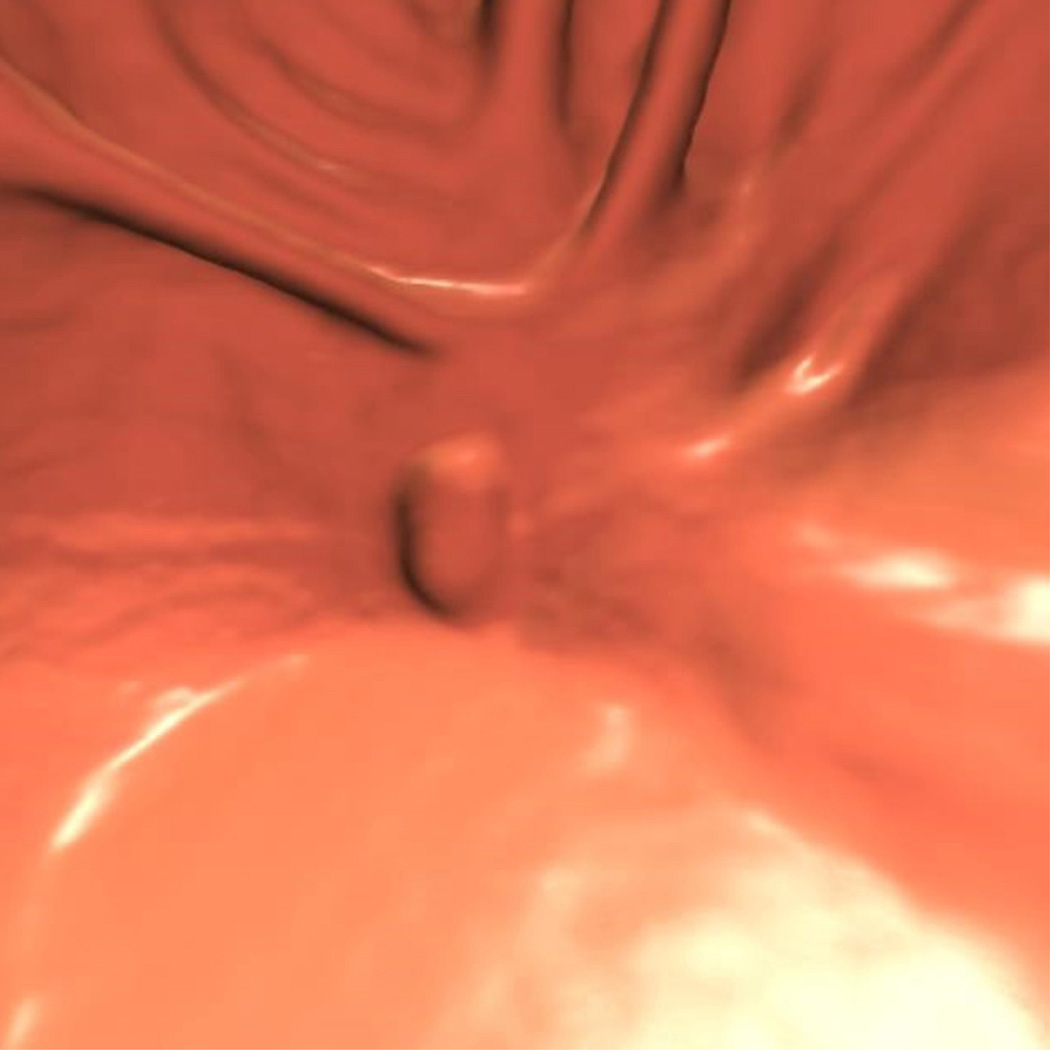
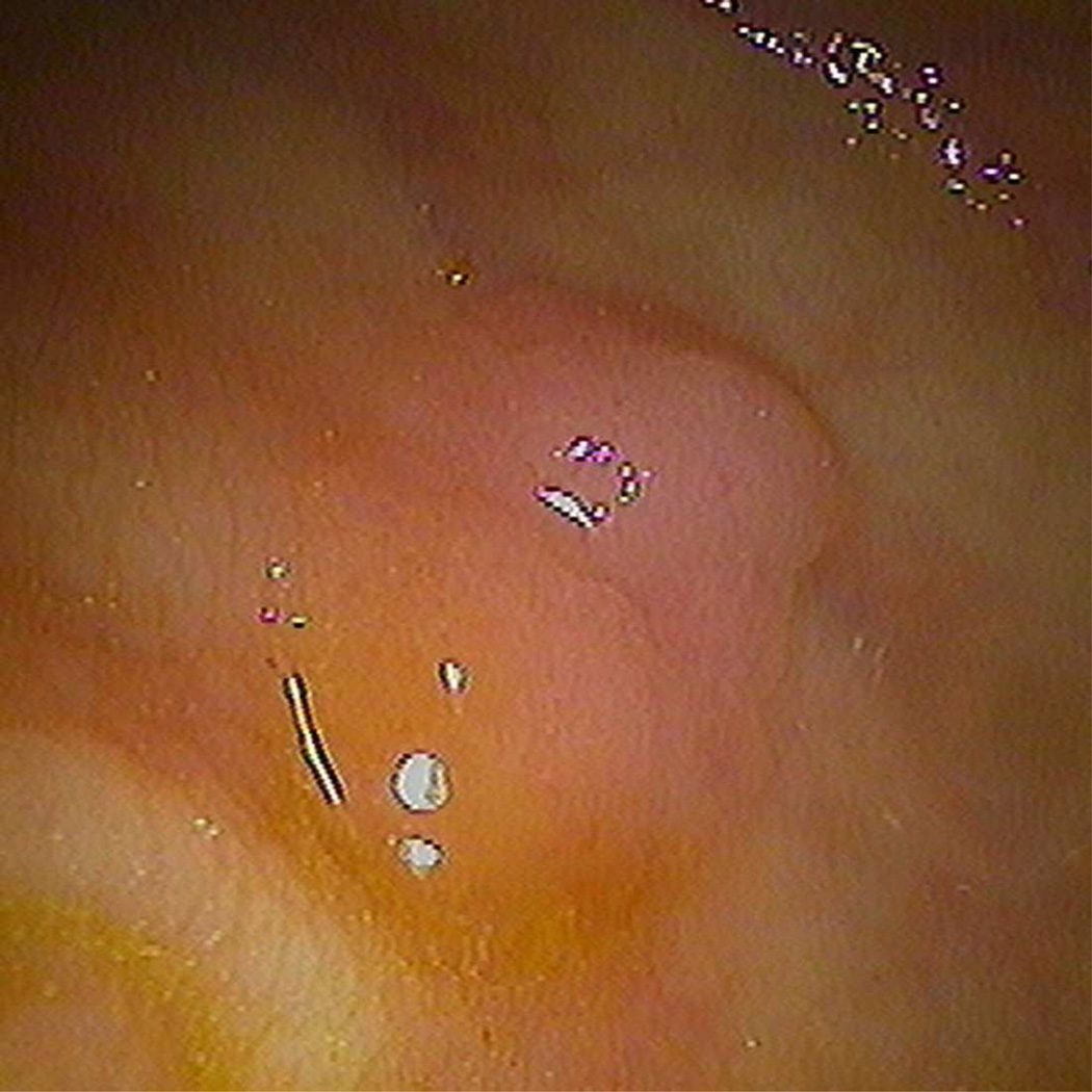
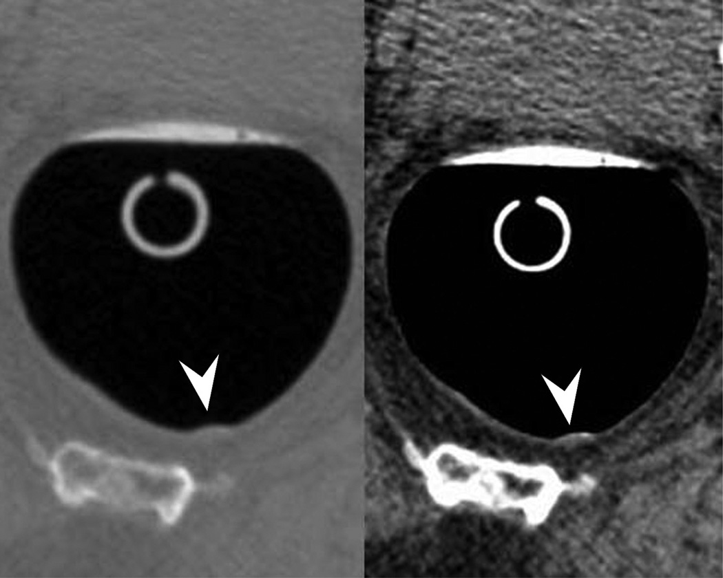
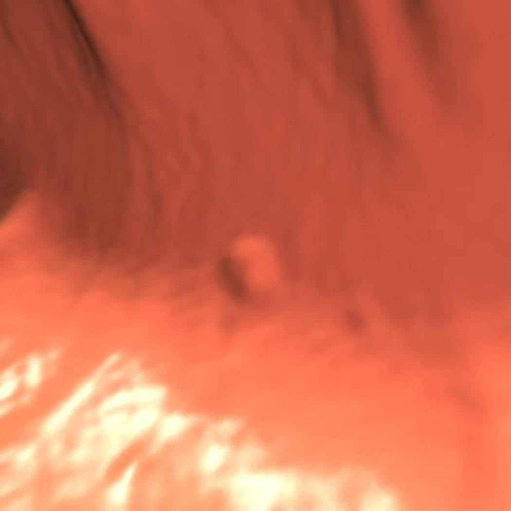
Figure 2.
Typical appearance of coated flat polyp. 2D supine images (A) in polyp (2000/0) and soft tissue (400/50) windows show a typical coated polyp (arrowhead) in the cecum. Note the subtle soft tissue thickening deep to the contrast coat, which is more evident on the soft tissue windows. 2D prone images (B) again demonstrates a focal contrast plaque of typical thickness overlying the polyp (arrowhead). The polyp is more prominent at 3D (C) as it represents a combination of polyp and contrast from this perspective. Optical colonoscopy (D) confirms the polyp which was hyperplastic in nature. 2D image (E) in another patient shows a thin film on contrast (seen in a quarter of coated polyps) (arrowhead). Note the polyp is less prominent on the 3D view (F).

