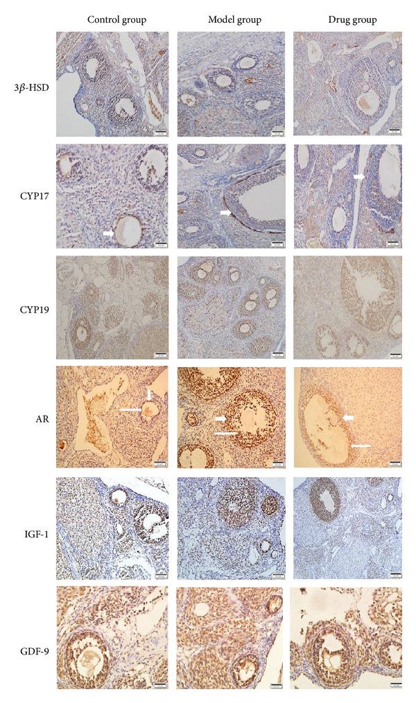
Figure 5.

Immunohistochemical staining of 3β-HSD, CYP17, CYP19, AR, IGF-1, and GDF-9 in three groups. As is shown above, CYP17 was primarily localized in theca cells (short thick arrows), AR was primarily expressed both in theca cells (short thick arrows) and in granulosa cells (long thin arrows), while the expressions of other proteins were not obvious.
