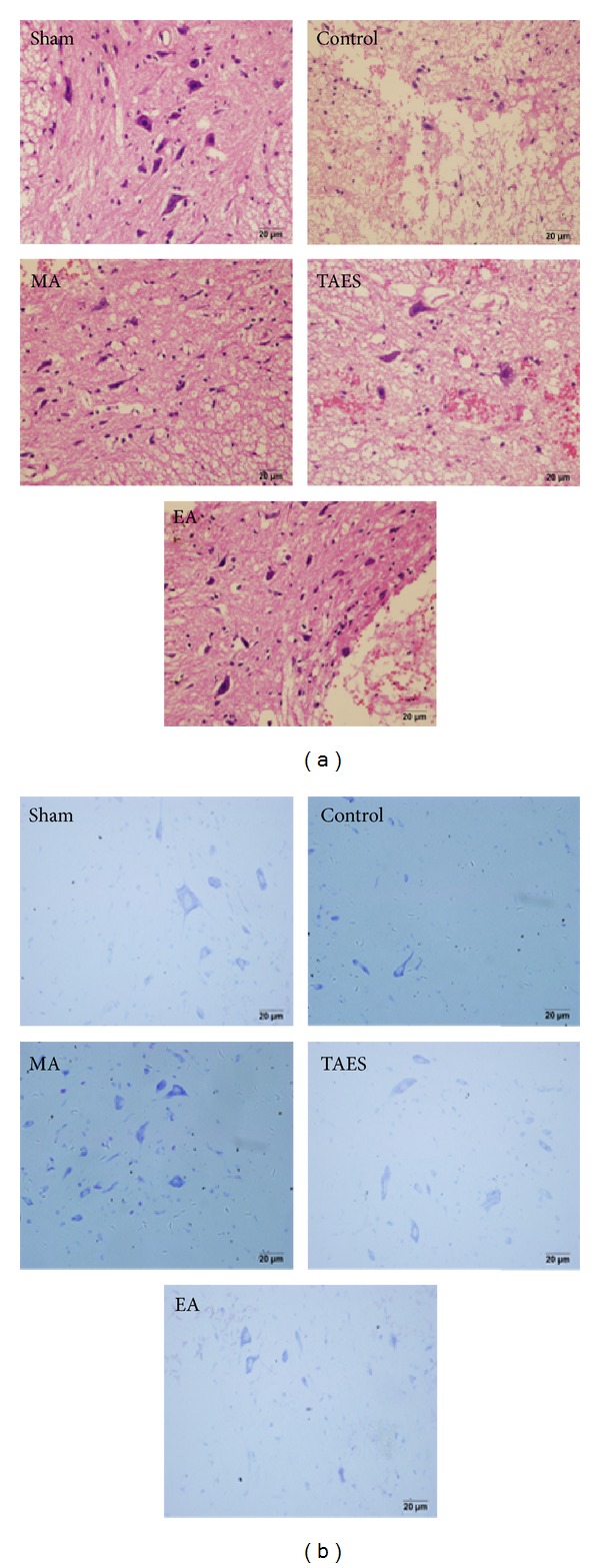
Figure 3.

HE staining and Nissl staining. (a) HE staining showed normal neural morphology in the sham group. In the control group, impacted spinal cord exhibited typical necrosis showing as broad hemorrhage, edema, and neuronal apoptosis with condensed nuclei. All in the EA, MA, and TAES group neurons displayed normal morphology with clear boundaries. Compared with the control group, hemorrhage and edema occurred in the EA, MA, and TAES groups. (b) In the sham group, neurons exhibited a large amount of densely stained toluidine blue granules in the cytoplasm. However, in the control group, the Nissl bodies dramatically decreased or even disappeared in the neurons. In the EA, MA, and TAES groups the quantity of Nissl bodies was restored compared with that of the control group and displayed patch morphology.
