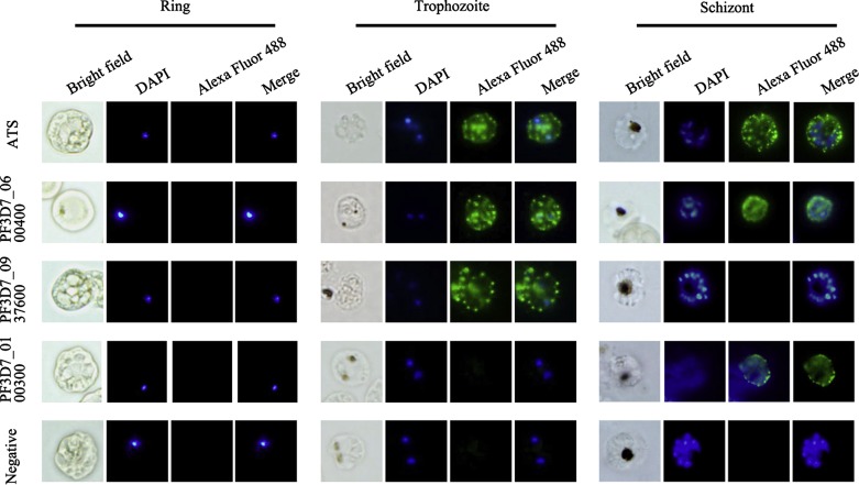
Figure 4.
Immunofluorescence assays with variant-specific antibodies. An immunofluorescence assay (IFA) was performed using anti-PF3D7_0100300-His, anti-PF3D7_0600400-His, and anti-PF3D7_0937600-His IgG (1:50) as primary antibodies and anti-ATS IgG as the control. Specific staining of infected erythrocytes (IEs) is observed as punctuated fluorescence patterns over the IE surface using a secondary antibody labeled with Alexa 488 (green). The pre-immune sera of the immunized rats did not result in any surface reactivity with the IE. DAPI (5 μg/mL) staining of DNA in the nuclei is blue. The anti-ATS IgG and anti-PF3D7_0600400-His IgG stained IEs of both the trophozoite and schizont stages, the anti-PF3D7_0937600-His IgG only reacted with IEs of the trophozoite stage, while the anti-PF3D7_0100300-His IgG only reacted with IEs of the schizont stage.

