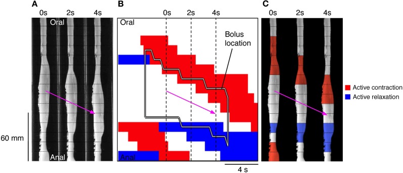
Figure 10.
(A) Using the same panel as shown in Figure 8 we have now added an outline of the bolus (black line shape in B) and the active states of neurogenic activity associated with the bolus movement. In (B) the red indicates active contraction at the oral end of the bolus, and blue indicates an active relaxation at the anal end of the bolus. The vertical hatch lines in (B) indicate the location of the silhouettes shown in (A). These active states are then superimposed upon these three silhouettes (C). The magenta arrow indicates the direction of the bolus movement. As can be seen in (C) the bolus movement is associated with active contraction (red) at the oral end of the bolus and active relaxation (blue) at the anal end.

