
Fig. 4. Nrf2-deficient mice exhibit more pronounced neuronal cell loss after retinal I/R injury.
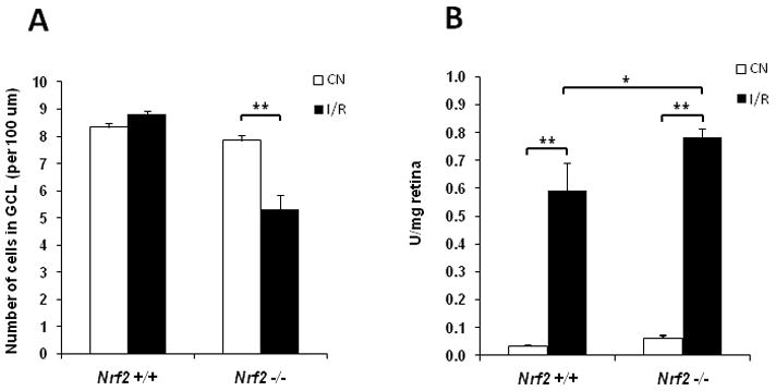
(A) Ganglion cell layer counting was performed as described in Methods. The number of cells in ganglion cell layer (GCL) of the I/R retina was significantly less than that of control retina (CN) in Nrf2 −/−mice 2 days after I/R injury, while no significant change was observed in the wild-type mice (n = 5, ** p < 0.01). (B) Apoptotic DNA Cleavage ELISA was performed 48 hrs after I/R injury. Nucleosomal DNA fragmentation was significantly increased in wild-type mice after I/R injury. There was a further increase in apoptotic DNA cleavage in Nrf2 −/− mice after I/R injury, beyond what was observed in wild-type mice. n = 5, * p < 0.05; ** p < 0.01.