
Figure 5.
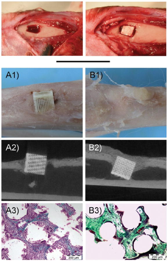
Photograph showing the Implantation of Ceramic Scaffolds with BMP-2 in rabbit bone. Top, images of the surgical procedure. (A) control scaffolds, (B) BMP-2 adsorbed scaffolds. (1) Gross appearance of harvested samples, (2) Representative mCT slides, (3) Representative histological images (Massons tricrome stainings). Reprinted with permission from [155], A. Abarrategi, et al., PLoS ONE 7, 34117 (2012). © 2012, PloS ONE.