
Figure 3.
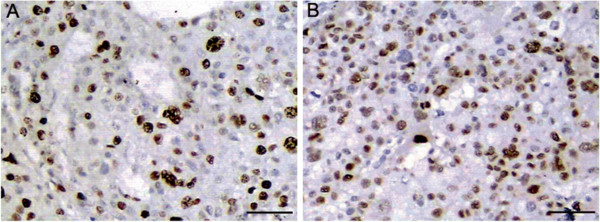
Immunohistochemical expressions of ki-67 and cyclin D1 in consecutive sections of an HCC tissue. Positive immunostaining of (A) ki-67 and (B) cyclinD1 were localized in the nucleus of cancer cell. Scale bars = 50 μm.
Official websites use .gov
A
.gov website belongs to an official
government organization in the United States.
Secure .gov websites use HTTPS
A lock (
) or https:// means you've safely
connected to the .gov website. Share sensitive
information only on official, secure websites.
Immunohistochemical expressions of ki-67 and cyclin D1 in consecutive sections of an HCC tissue. Positive immunostaining of (A) ki-67 and (B) cyclinD1 were localized in the nucleus of cancer cell. Scale bars = 50 μm.