
Figure 2.
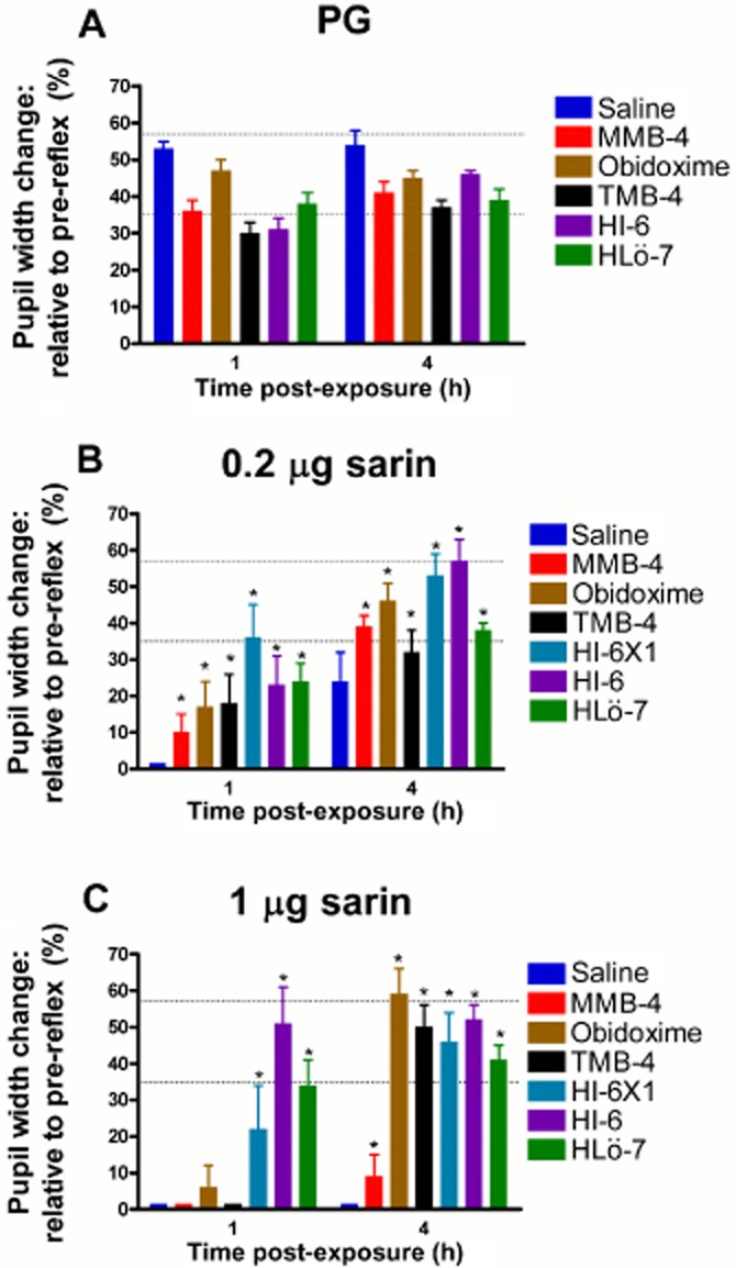
Effect of oxime treatments on the pupillary light reflex following sarin exposure. Treated eyes, as described in Figure 1 (two drops of topical 2.5% oxime), were illuminated with 350 lux for 2–3 s, 1 and 4 h following exposure to PG (A), 0.2 μg (B) or 1 μg (C) sarin. Each point is presented as % change relative to the pre-reflex pupil width and represents the mean ± SEM of 12 animals. Normal light reflex range is indicated by the dotted horizontal lines. Differences between treatments following exposure of 0.2 or 1 μg sarin at both time points post-exposure are statistically significant (by anova), at a level of *P < 0.01 (vs. saline at the same time point).