Figure 1.

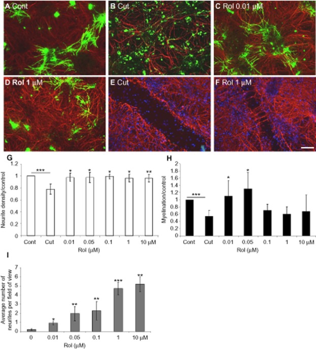
Rolipram induces myelination and neurite outgrowth. Immunolabelling of control, (A), cut (B: adjacent to the lesion, E: lesion site), and cut cultures treated with 10 nM (C) or 1 μM (D: adjacent to the lesion, F: lesion site) rolipram (Rol) with SMI-31 (red) and anti-PLP/DM20 (green) antibody. Cultures were cut at day 24, and treated with varying concentrations of rolipram (μM) 1 day after cutting. The cultures were treated for 6 days prior to immunocytochemistry and quantification of neurite density (G) and myelination (H) surrounding the lesion, as well as neurite outgrowth into the lesion (I). Scale bar 100 μm. *P < 0.05, **P < 0.01, ***P < 0.001, significant differences between control and cut, and treatments and cut.
