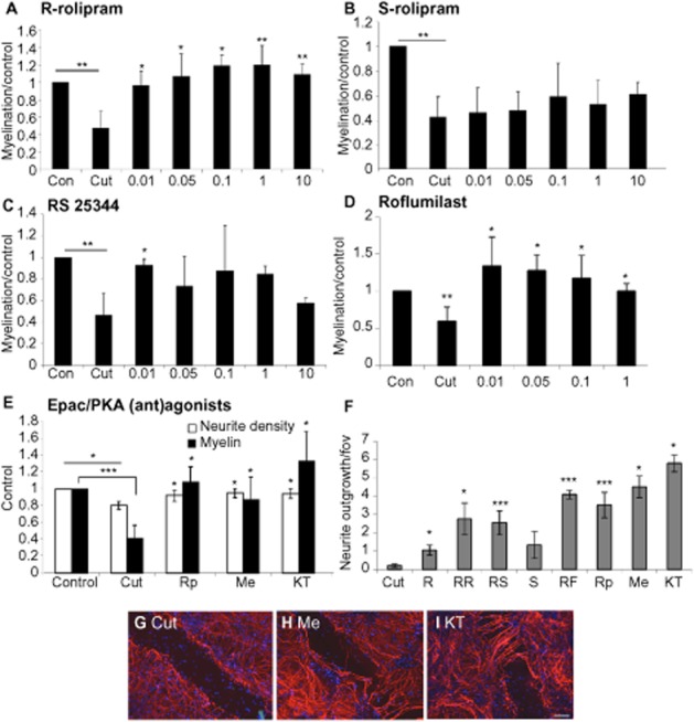
Figure 3.

Myelination is primarily mediated via HARBS and Epac. Cut myelinating cultures were treated with varying concentrations (in μM) of R-rolipram (RR; A), S-rolipram (S; B), RS25344 (RS; C),roflumilast (RF; D), Epac/PKA (ant)agonists RpcAMPS (Rp), Me-cAMP (Me), KT5720 (KT; E), and the change in myelination surrounding the lesion, normalized to control, quantified. Neurite density surrounding the lesion was restored to control levels by all drugs tested (data not shown). In addition, the extent of neurite outgrowth into the lesion is shown in F (all at 10 nM), and representative images of the lesion site are shown of untreated cut cultures (G), cultures treated with Me-cAMP (Me; H) and KT5720 (KT; I). Scale bar 100 μm. *P < 0.05, **P < 0.01, ***P < 0.001; significant differences between control and cut, and treatments and cut.
