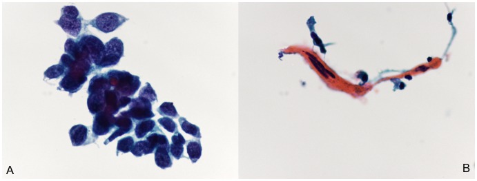
Figure 1. Cytomorphology of lung squamous cell carcinoma (SCC) using ThinPrep bronchial brushing cytology.

Small clusters of malignant cells with features of poorly differentiated carcinoma. (A), Well-differentiated SCC. Cytological features are characterized by individual cells or cohesive sheets of tumor cells with clear cell borders and a dense cytoplasm. (B), Poorly differentiated SCC. Cytology shows high cellularity and small groups of tumor cells. The nuclear/cytoplasmic (N/C) ratio was usually high (Papanicolaou staining; the original magnification was 400×).
