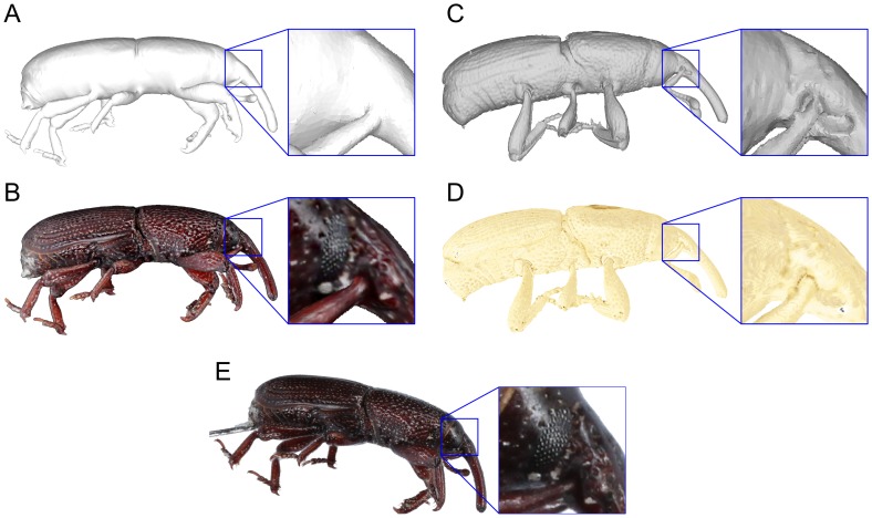
Figure 10. Comparison of a natural-colour 3D model, a Micro CT reconstruction and 2D image at a similar angle.
The surface geometry of the natural-colour 3D model (A) is less detailed than the Micro CT model (C) and missed concavities such as the antenna socket shown in the enlarged inset of C. However, the natural-colour 3D model can capture useful surface information such as the compound eye in the enlarged insect of B. False-colour Micro CT model (D) and a 2D image (E) are shown for comparison.

