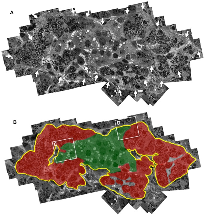
Figure 1. Demonstration of pancreatic acinar-like cell clusters touching islet-cell clusters that are covered by a common capsule.
A: Continuous basement membranes (BMs) and extracellular matrix (ECM) (arrows) cover the two cell clusters. A combined figure of 65 electron microscopic photos is shown. B: Schematic demonstration of Figure 1A. The yellow line indicates continuous BMs and ECM surrounding islet cell (green) and acinar-like cell (red) clusters. LB: lipofuscin body.

