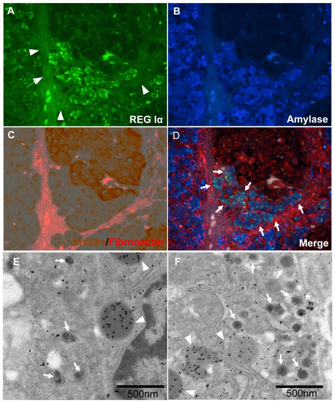
Figure 5. REG Iα-positive cell cluster contacting with beta cell cluster and surrounded by common BMs and ECM.
A–D: Immunostaining for REG Iα (green) (A), acinar-like cell marker, amylase (blue). (B), and double immunostaining with insulin (brown) and fibronectin (red) (C). Auto-fluorescence of collagen fiber surrounding islet is observed (A, arrowheads). The merged image (D) shows that amylase-positive acinar-like cells that are in contact with beta cells express REG Iα protein (light blue: arrowheads), and the acinar-like cell cluster is surrounded by BMs/ECM (red, fibronectin). E: Electron-immunostaining with immuno-gold for REG Iα (20 nm: arrowheads) in acinar-like cell touching a beta cell containing insulin (5 nm: arrows). REG Iα is mainly localized in the center of the vesicle that is near the beta cell wall. F: Immuno-electron microscopy with immunogold for REG Iα (25 nm: arrowheads) and insulin (5 nm: arrows). Densely stained REG Iα vesicle (*) is just beside the cell wall touching a beta cell. Dissolved vesicles positive for REG Iα (arrowheads) are observed in insulin-containing beta cells.

