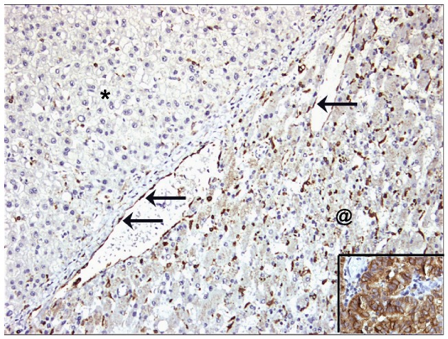
Figure 2. COX-2 immunostaining on HCC (upper left, marked with *) and adjacent liver parenchyma (lower right, marked with @).
COX-2 expression is present on endothelial cells (arrows) at the interface between HCC and non-tumorous liver parenchyma. In the liver parenchyma COX-2 expression is mainly present in Kupffer cells and some inflammatory cells. Note the absence of staining in hepatocytes and tumor cells. The insert (lower right hand corner) shows COX-2 immunostaining on coloncarcinoma as a positive control.

