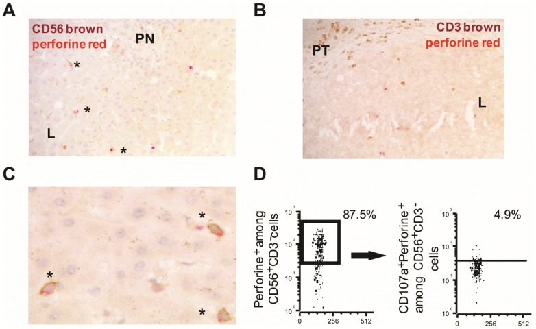
Figure 3. Immunohistochemical detection of perforin+CD56+ and perforin+CD3+ IH cells from HCV-infected patients and flow cytometric analysis.
Representative pictures from immunohistochemical staining of HCV-infected liver (n = 4) with double perforin/CD56 labeling (A) showing perforin+CD56+ cells characterized by brown CD56+ staining and red cytoplasmic perforin+ staining in HCV-infected liver with septal fibrosis and low A1 Metavir activity (magnification ×20). Many double positive CD56+perforin+ cells (asterisk) were detected, mainly localized in lobules (L), far away from piecemeal necrosis (PN). B) Detection of brown CD3+ cells and red perforin+ cells in HCV-infected liver with septal fibrosis. Rare double positive CD3+perforin+ cells were detected in lobules. C) High magnification (x40) showing that perforin granules were polarized at the apical pole of IH-CD56+ cells. D) Flow cytometric analysis depicting perforin+cells gated on CD56+CD3− cells and CD107a+ gated on perforin+NK cells.

