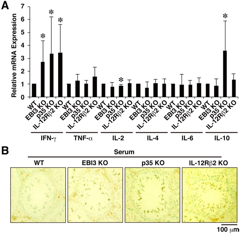
Figure 3. Augmented IFN-γ expression and autoantibody production in mice deficient in EBI3, p35, and IL-12Rβ2.
(A) Total RNA was prepared from testes of WT mice and deficient mice (12 weeks old; n = 5 per group) and subjected to real-time RT-PCR using primers for various cytokines. Ratio of relative expression level of each cytokine in deficient mice to that in WT mice was calculated. Data are shown as mean ± SD. *P<0.05 compared with WT mice. (B) For detection of serum autoantibodies, cryostat sections of testes of WT mice (12 weeks old) were immunohistochemically stained with diluted serum samples (×50, n = 5 per group) obtained from WT mice and deficient mice (12 weeks old). The sections were also counterstained with methyl green. Representative histology images are shown. Positive cells are shown as dark brown spots. Similar results were obtained in three independent experiments.

