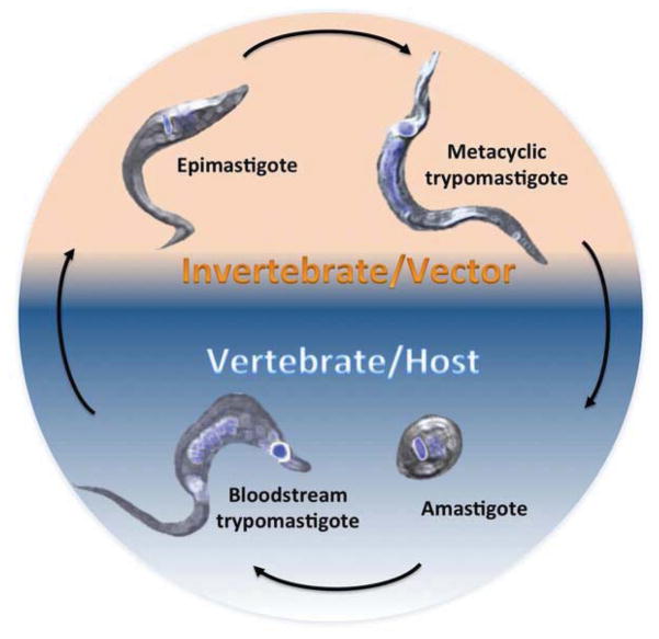
Fig. 1. T. cruzi life cycle.
Epimastigotes and metacyclic trypomastigotes develop in the gut of the insect vectors. In the vertebrate host, the developmental stages are circulating bloodstream trypomastigotes and amastigotes. In blue are stained the nucleus and the kinetoplast (the single mitochondrion of T. cruzi). The shape and relative position of both organelles changes through the development of the parasite and is one of the hallmarks of each developmental stage. Background colors indicate the stages found on each host, orange for parasite forms developed in the insect vector and blue for those found in mammalian hosts.

