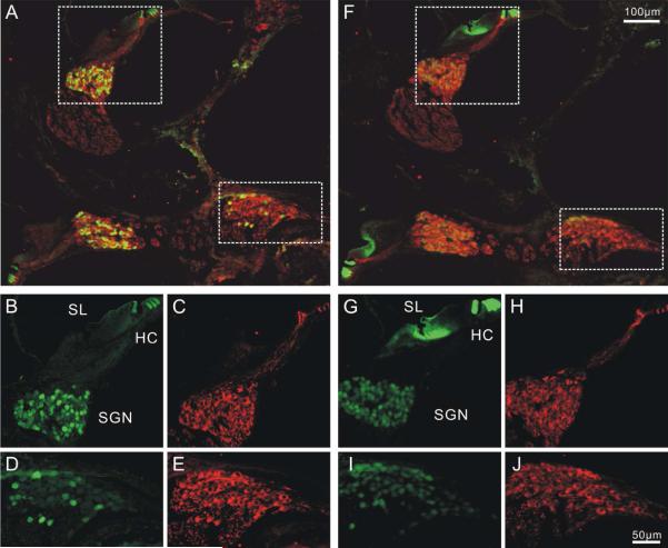
Figure 3. Distribution patterns of calretinin and calbindin in P6 mouse cochlea.
Two cochlear sections in close proximity were stained with anti-calretinin (A-E) and anti-calbindin antibodies (F-J) respectively. Both anti-calretinin and anti-calbindin antibody staining showed heterogeneous patterns in the spiral ganglion. HC, hair cell. SGN, spiral ganglion. SL, spiral limbus. A, Low magnification image of cochlear sections double labeled with anti-β-tubulin (red) and anti-calretinin (green) antibodies B-E, High magnification images of middle (B-C) and basal (D-E) neuronal regions enclosed by dotted line in A. F, low magnification image of a cochlear section showing double labeling of anti-calbindin (green) and anti-β-tubulin (red) antibodies. Prominent calbindin staining was also observed in the spiral limbus G-J, High magnification image of the middle (G, H) and basal (I,J) regions as squared in F. Scale bar in F applies to A, F. Scale bar in J applied to B-E, G-J. A magenta-green version of this figure is available as supplementary materials.

