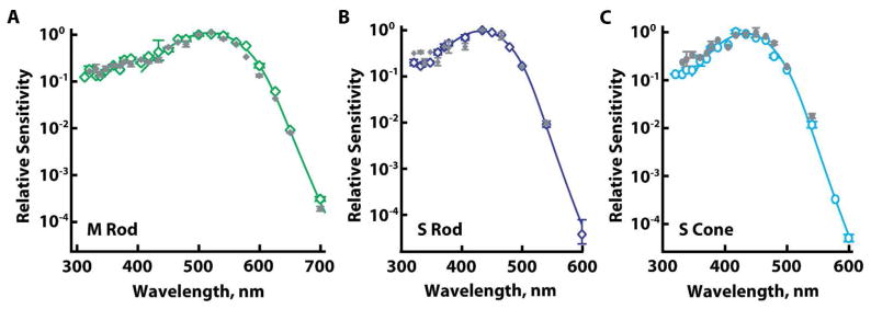
Figure 5.

Lack of physiological evidence for opsin mixtures in rods and in S cones. Averaged, dark-adapted, spectral sensitivities (open symbols) were unchanged by partial bleaching (filled symbols). Solid lines are template fits to dark-adapted results. A. M rods (n=5) exposed to 560 nm light that bleached 85 ± 3% pigment. B. S rods (n=3) exposed to 450 nm light to bleach 94 ± 2% pigment. C. S cones (n=2) exposed to 450 or 465 nm to bleach 86 ± 11% pigment. Error bars represent SEM.
