Figure 7.

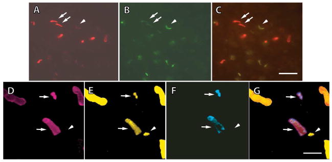
Immunohistochemical evidence for the coexistence of multiple visual pigments within UV cones and L cones of larval salamanders. A–C. Staining with Blue-N, an antibody to S opsin (A), and UV-N, an antibody to UV opsin (B); Blue-N was conjugated with Alexa Fluor 594, and UV-N with Alexa Fluor 488. Blue-N labeled some outer segments brightly (arrows) and others dimly (arrowhead), while UV-N labeled the same outer segments in a reverse pattern, indicating that some cones contained a mixture of S and UV visual pigments. The composite image is shown in (C). D–G. Retina triply labeled with CERN956 for L opsin (D), TA2 for cone transducin α-subunit (E), and UV-N (F); all four images are of the same field with the composite shown in G. CERN956 and UV-N were conjugated with Alexa Fluor 594 and Alexa Fluor 350, respectively; TA2 was biotinylated and visualized with a FITC-conjugated secondary. Some outer segments were positive for L and UV opsins (arrows). One cone outer segment showing TA2 immunoreactivity belonged to an S cone (arrowhead). Scale bar = 16 μm in A–C, and 8 μm in D–G.
