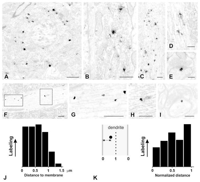
Figure 6.
Electron micrographs of pre-embedding labeling for IRSp53. All images are from layers II–III of S1 neocortex; A–E are from immunoperoxidase material; F–I are from silver-enhanced nanogold. A, patches of nickel-intensified DAB immunoreaction in the soma of a small pyramidal cell. Label is in cytoplasm, excluded from nucleus. B, higher magnification image from another cell shows reaction product associated with rough endoplasmic reticulum and Golgi apparatus. C, large proximal dendrite contains numerous patches of immunoreaction. D, immunoreaction is associated with microtubules in a thin dendritic shaft. E, small patch of immunoreaction at the cytoplasmic fringe of the PSD of a dendritic spine. F, pre-embedding silver-enhanced immunogold shows immunolabel associated with microtubules (boxed area on left, enlarged in G) and with tubulovesicular structures (boxed area on right, enlarged in H). I, high-magnification image shows silver-enhanced gold particle lying close to the PSD in a small dendritic spine. J, histogram shows distance of gold particles to the plasma membrane (N = 161 particles from ten micrographs of randomly-selected dendritic shafts). K, histogram shows the same data, replotted to show “normalized distance” such that 0 corresponds to membrane, and 1 to center of dendrite (see inset on left).
Scale bar = 1 μm in A; 500 nm in B, C, D; 200 nm in E, 500 nm in F, G, H; 200 nm in I.

