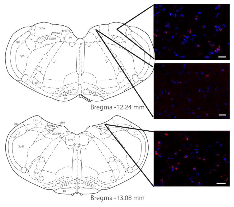
Figure 2.
C-Fos protein (red) visualized by immunofluorescence staining of vestibular neurons activated using low frequency sinusoidal galvanic vestibular stimulation (sGVS). Activated vestibular neurons are illustrated at two levels of the vestibular nuclear complex (VNC), and in both the spinal and medial vestibular nuclei (SpVN and MVN, respectively). DAPI (blue) was used as a marker for neuronal nuclei. As previously reported (Holstein et al., 2012), c-Fos-positive vestibular neurons were observed in the caudal half of SpVN and throughout the non-magnocellular MVN after sGVS stimulation. Atlas templates were obtained from (Paxinos and Watson, 2005). Scale bars: 20 μm.

