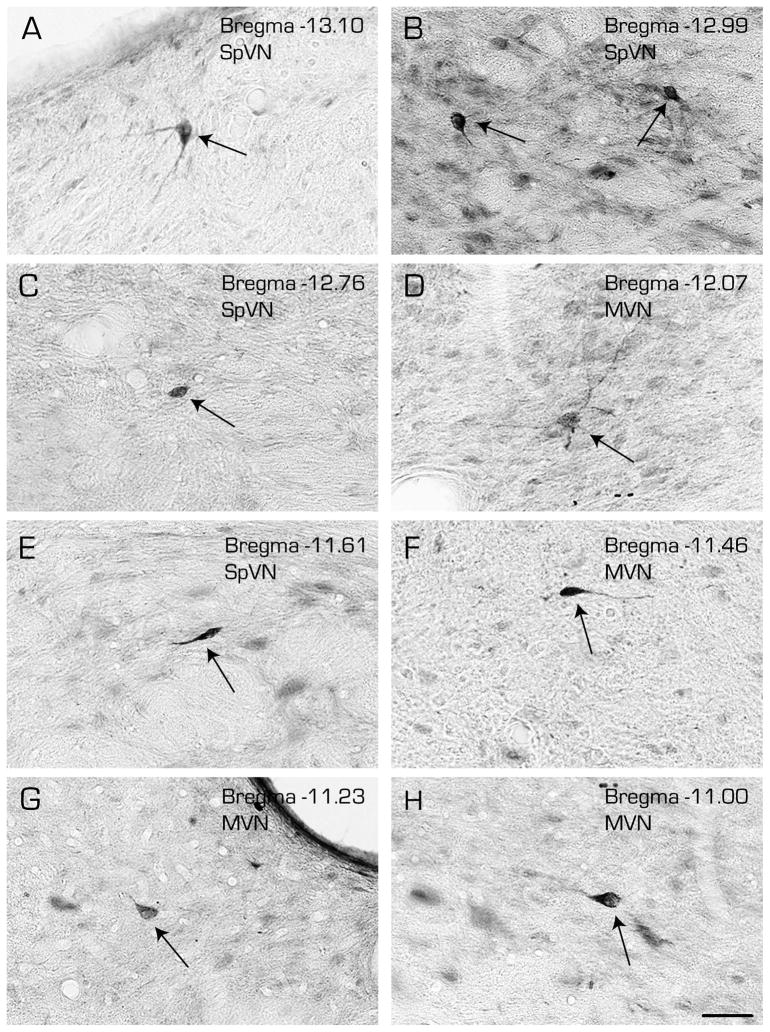
Figure 6.
Immunoperoxidase/diaminobenzidine-stained Vibratome sections through the caudal vestibular nuclei illustrating the three morphological types of vestibular neurons that were retrogradely-filled following a FluoroGold tracer injection into CVLM. Multipolar (A, B-right, D), globular (B-left, C and G) and fusiform (E, F and H) cells (arrows) were observed throughout the caudal half of SpVN and through the caudal and parvocellular MVN. Estimated Bregma levels are based on matching anatomical boundaries with atlas drawing from (Paxinos and Watson, 2005). Scale bar in H: 50 μm for all panels.

