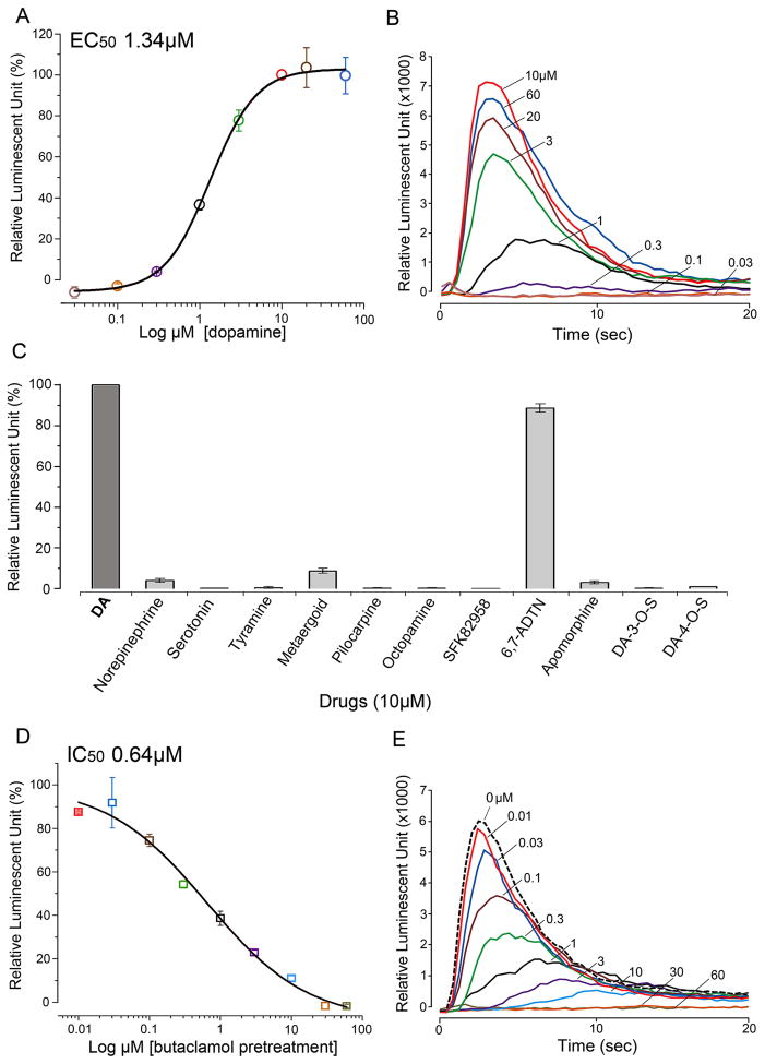
Figure 2.
Bioluminescent aequorin reporter assays for InvD1L receptor expressed in Chinese hamster ovary cells (CHO-K1). (A) Dose-response curve of InvD1L receptor to various doses of dopamine (DA). (B) Representative luminescence responses of InvD1L when treated with different doses of DA (30 nM to 60 μM). (C) Agonistic activities of various chemicals (10 μM) on the InvD1L receptor. (D) Dose-responses curve of the antagonistic activity of (+)butaclamol on the DA-mediated (10 μM) InvD1L receptor activity. (E) Representative luminescence responses of InvD1L to 10 μM DA after pre-incubation with different concentrations of (+)butaclamol for 15 minutes. For details of chemicals used, see section 2.1 Tick samples and chemicals. The colors in A and D correspond to those of the curves in B and E, respectively. The bars in A, C and D indicate the standard error for three replicates.

