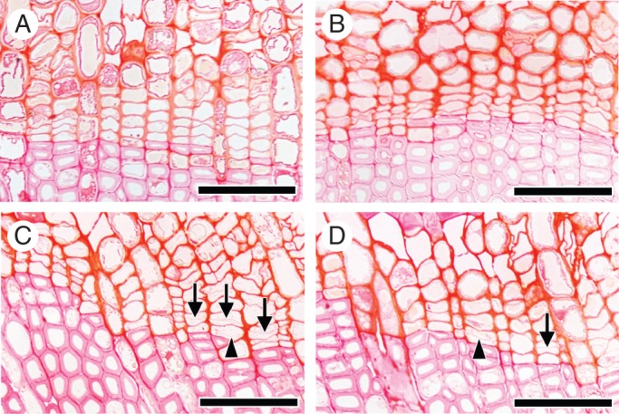
Fig. 3.

Light micrographs showing transverse views of the cambial zone after 41 d on 13 March 2012. In the control (A) and the disbudded seedlings (B), there were no new thin cell plates in the cambial zone. In contrast, in the heated portion of a heated seedling (C) and of a heated plus disbudded seedling (D), new thin cell plates were visible in the cambial zone. These new thin cell plates were observed in the first layer (arrowheads; C and D) and second or third layer (arrows; C and D) of fusiform cambial cells from the previous year's xylem. Division of cambial cells was observed in the heated portion of stem. Scale bars = 50 μm.
