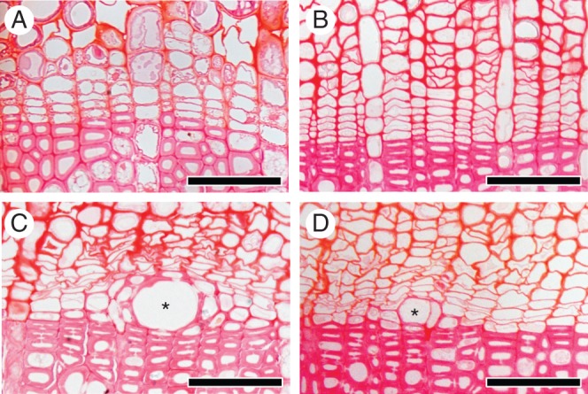
Fig. 4.

Light micrographs showing transverse views of the cambial zone after 55 d on 27 March 2012. In the control (A) and disbudded seedlings (B), there were no new thin cell plates in the cambial zone. In the heated portion of the heated seedlings (C) and of a heated plus disbudded seedling (D), vessel elements with deposition of secondary walls were visible (asterisks; C and D). Scale bars = 50 μm.
