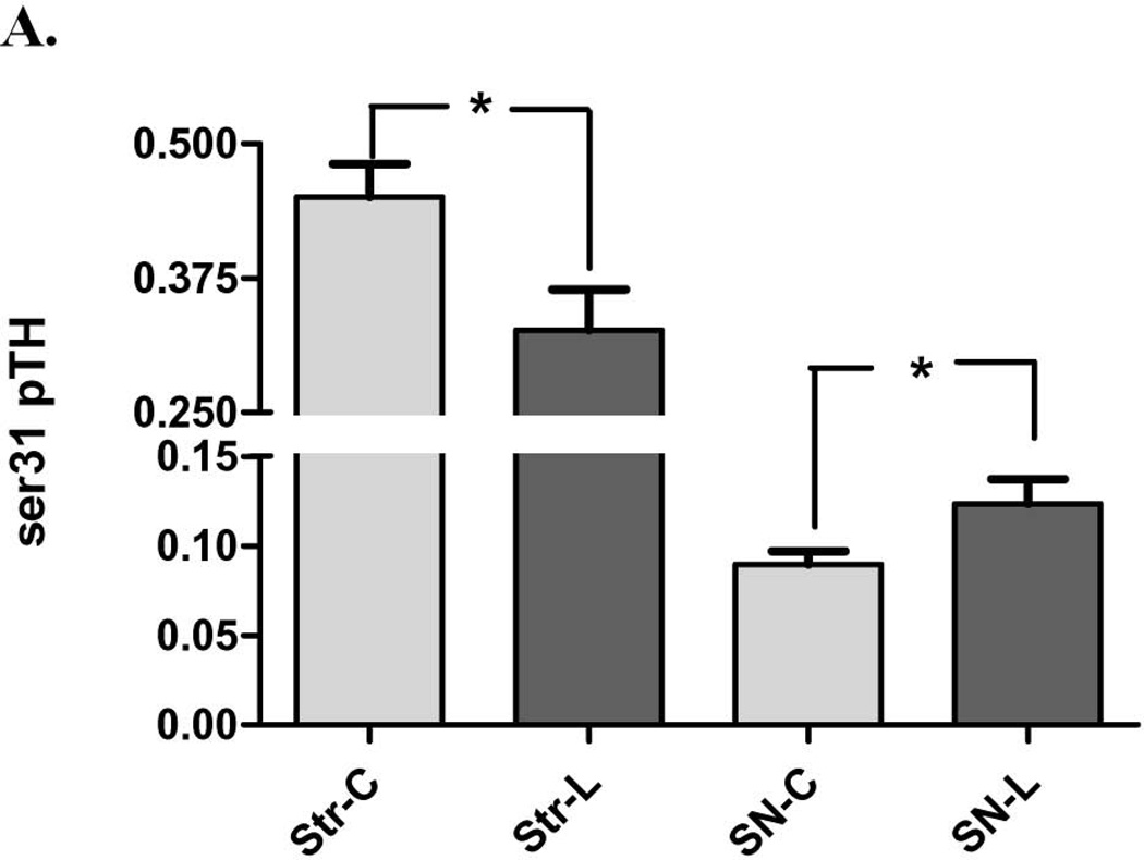
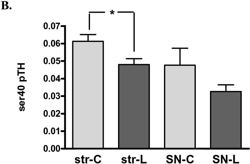
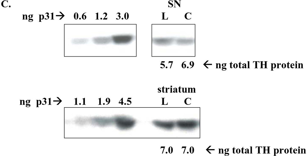
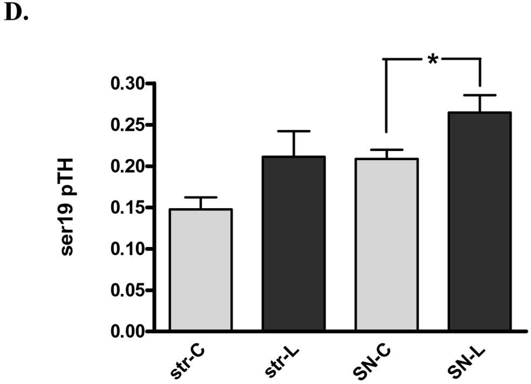
Figure 6. Impact of 6-OHDA lesion on TH phosphorylation stoichiometry. A. ser31 TH phosphorylation in striatum (str) vs. substantia nigra (SN).
Ser31 decreased on average 28% following 6-OHDA lesion in striatum (n=11, *p=0.0195, t=2.78, df=10) but increased 37% in SN (n=11, *p=0.0125, t=3.04, df=10). B. ser40 TH phosphorylation in striatum (str) vs. substantia nigra (SN) Ser40 decreased on average 22% following 6-OHDA lesion in striatum (n=7, *p=0.011, t=3.65, df=6) but did not produce a consistent increase or decrease in the SN (n=5, p=0.27, t=1.32, df=3). C. representative western blot results from ser31 phosphorylation in the SN (top panel) and striatum (bottom panel). ng of phospho-ser31 loaded for standard curve is indicated and selected image from a lesioned (L) and contralateral control tissue (C) from the same rat in the same assay to show, with a comparable total TH protein load (~6 – 7 ng) that the phosphorylation at ser31 is increased in lesioned SN (represented pair not adjacent to standard curve), but decreased in lesioned striatum, compared to corresponding contralateral control tissue (represented pair adjacent to standard curve). D. ser19 TH phosphorylation in striatum (str) vs. substantia nigra (SN). Following 6-OHDA (SN-L), Ser19 phosphorylation (60 kDa band only) increased 27% in SN relative to control (SN-C) (n=7, *p=0.006, t=4.22, df=6). There was a trend (p=0.061) toward an increase in ser19 TH phosphorylation in the lesioned striatum. Note: results reflect 60 kDa immunoreactive band only for all measures.


