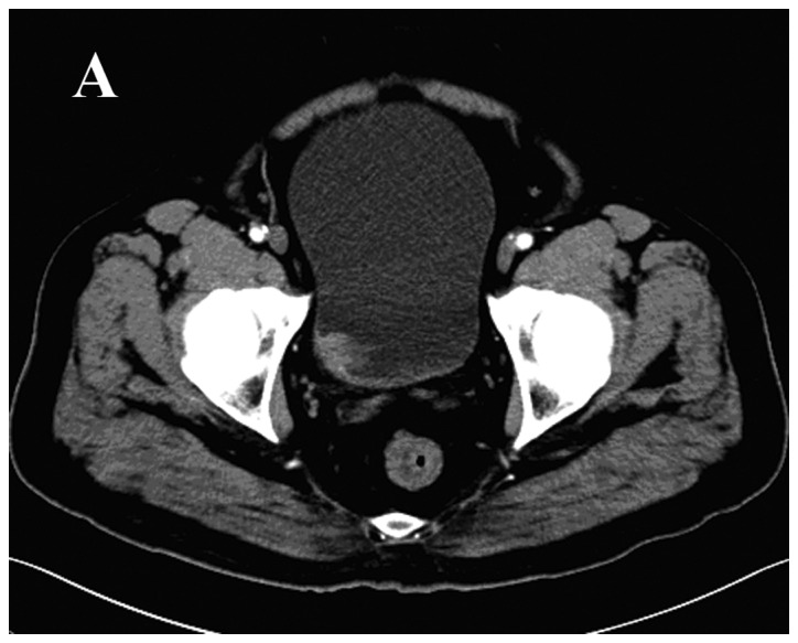
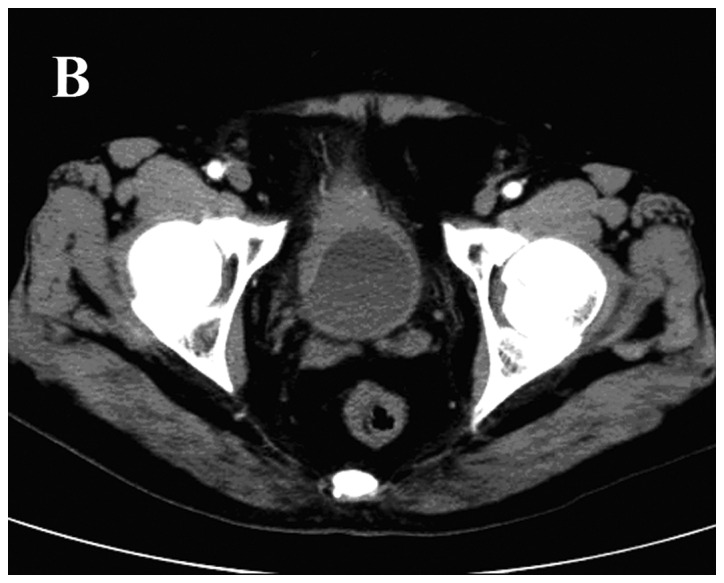
Figure 1.


Radiological features of the case. Arterial-phase computed tomography scan revealing (A) a 2.5 cm-diameter solid pedunculated mass at the right side of the bladder wall, and (B) thickened anterior and right bladder walls with significant mucous membrane enhancement. No significant bladder mass was detected.
