Figure 2.


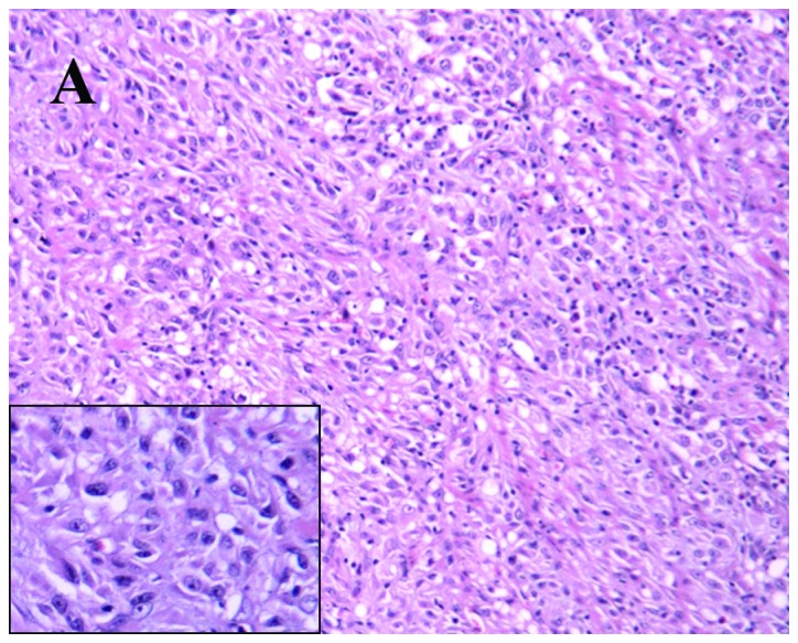
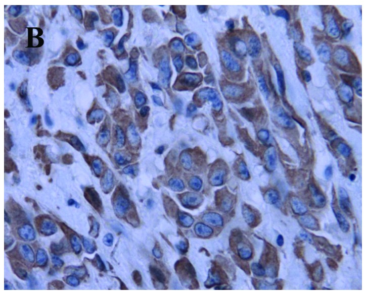
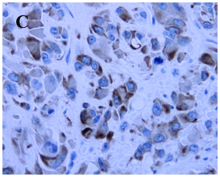
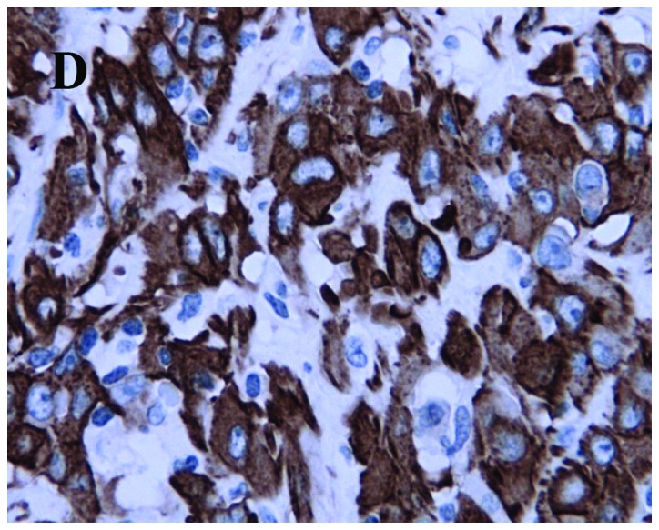
Histopathological and immunohistochemical features of the case: (A) Irregular fascicles of bland-looking spindle cells with several fascicles of chronic inflammatory cells scattered in myxoid stroma (hematoxylin and eosin staining; magnification, ×200); (B) vimentin is strongly expressed in the spindle cells (magnification, ×400); (C) positive cytokeratin immunohistochemical staining (magnification, ×400); and (D) focal positivity of smooth muscle actin staining (magnification, ×400).
