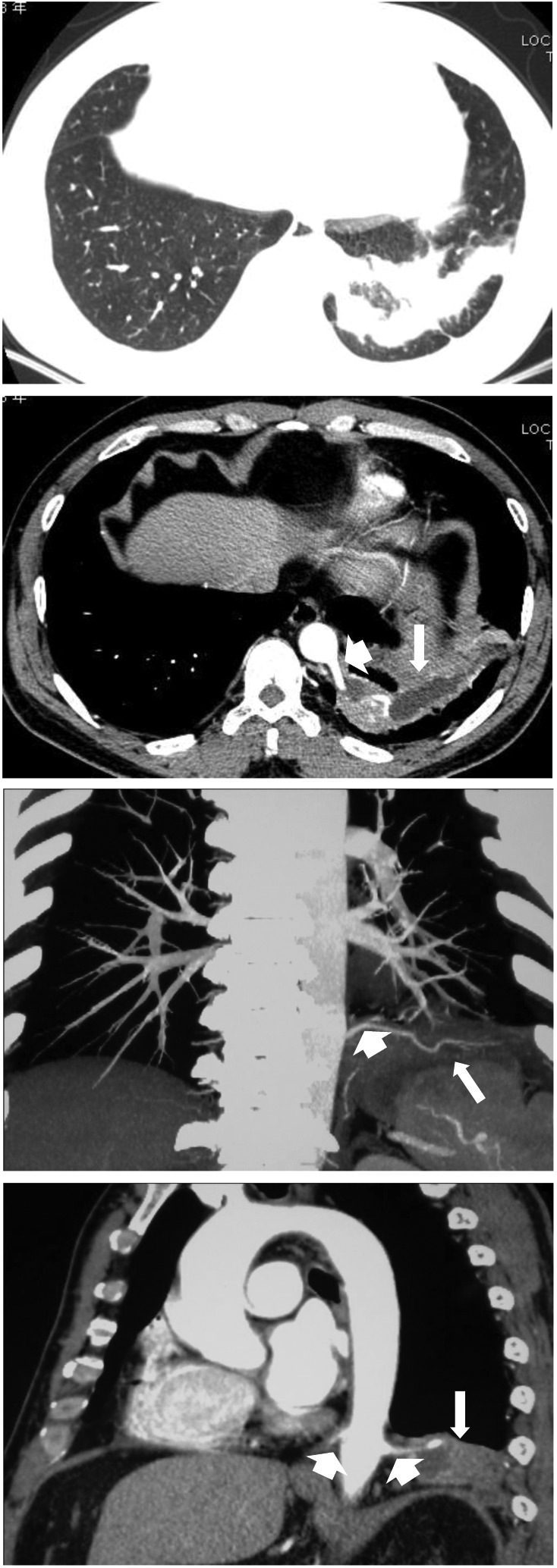
Figure 2.

Chest high-resolution computed tomography scanning and 3D image reconstruction showed a mass in the left lower lobe and two anomalous arteries (short arrow) arising from the descending thoracic aorta and extending into the sequestered lung (long arrow).
